Let’s talk about measurements. You are scanning and scrutinizing the organs for their echotexture, echogenicity, size, and shape and in doing so we are trying to determine normal or abnormal to give us some clue as to whether there is a disease process going on or not (remembering that some organs can appear normal sonographically but may still have disease). But we do know that disease processes can alter organ size. So, we have measurements that have been published for normal sizes of certain organs. Now the caveat to this is that there is no one concrete measurement for each organ instead there are ranges and vary depending on which publication you read.
With this blog my goal will be to give you a general reference measurement of the organs that will be the most pertinent to the general practitioner with the understanding that these are guidelines. Relying heavily on measurements is at best unreliable. One has to take into effect operator reliability and repeatability. Hopefully, you have purchased an ultrasound book and are using it to help you scan. Within the ultrasound book is the information on measurements and their parameters. I will list some of the ultrasound books available at the end of this blog. The measurements here are taken from these texts.
So, with that in mind let’s get started.
- Duodenum – measure the height from the edge of the serosa to the lumen
- Dog Up to 6 mm
- Cat 2 – 2.5 mm

- Gallbladder Wall
- Dog 2-3 mm
- Cat <1mm

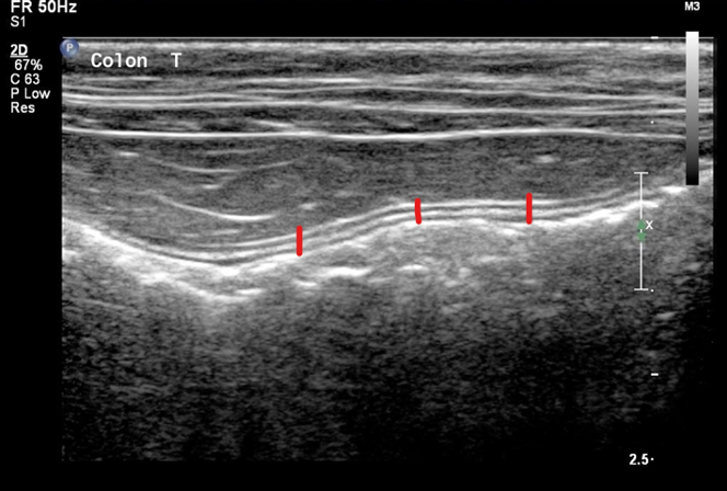
- Colon – measure height from serosa to lumen
- Dog 2 – 3 mm
- Cat 1 mm
- Jejunum measure as you would duodenum from serosal edge to lumen.
- Dog 2 – 5 mm
- Cat 2 – 2.5 mm
- Ileum
- Dog 2 – 4 mm This is not a normal ileum, could probably find a better example- hard to see wall layering here
- Cat 2.5 – 3.2 mm

- Gastric wall
- Dog 3 – 5 mm

-
- Cat 2 mm (inter-rugal)3,4 and 4 mm (rugal fold thickness)

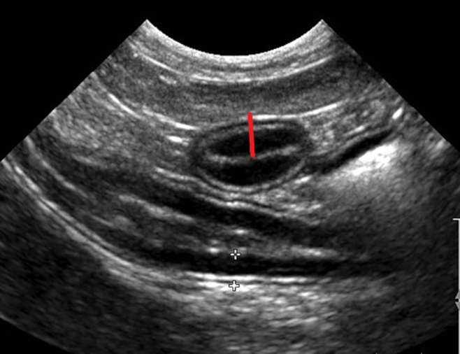
- Kidney – measured in the longitudinal/sagittal plane
- Dog Currently, there is no widely accepted method for determining ultrasonographically normal kidney size for dogs. Ultrasonographic size is usually subjective.
- Cat 3 and 4.3 cm

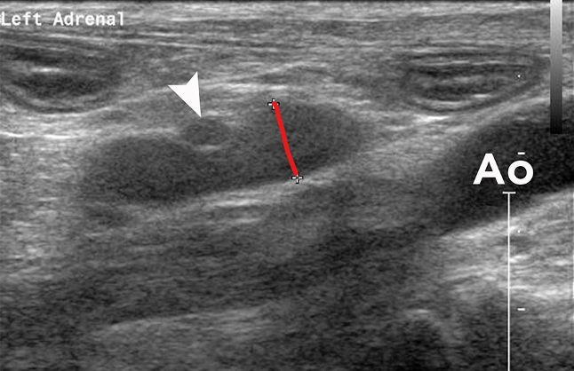
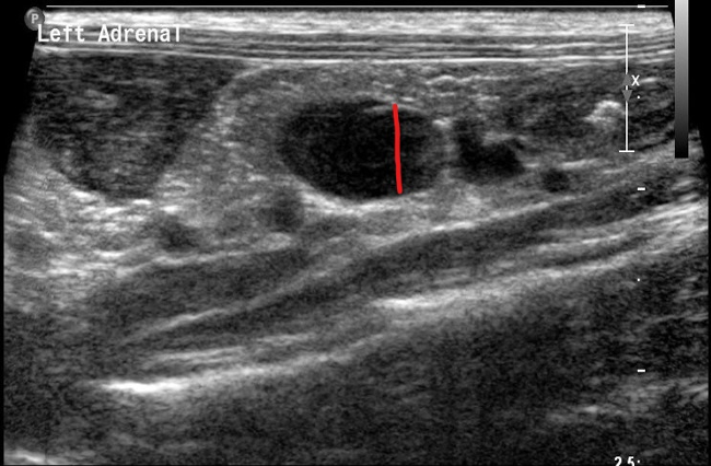
- Adrenal – measure at the caudal pole in longitudinal/sagittal plane
- Dog up to 7.4 mm
-
- Cat 3.7 – 4.9 mm
- Lymph nodes – measure in longitudinal/sagittal plane
- Dog and Cat < 5mm

- Prostate measure in the longitudinal/sagittal and transverse planes.The size and position of the normal canine prostate gland can vary with age, size, and intact or neutered status of the animal. In sexually immature or neutered dogs, the prostate is much smaller and often has a relatively hypoechoic and homogeneous appearance. Measurements for the prostate can be used to document the progression of disease or response to treatment.

There you have it. Some of the measurements that are pertinent. I gently remind you that measurements are guidelines and there is room for operator error. When in doubt refer to Ultrasound books and publications for guidance!
References: Nyland and Mattoon, D’Anjou and Penninck, Today’s Veterinary Practice, and Dr. Federico Vilaplana Grosso, LV, DECVDI, DACVR
Ultrasound Books and Materials
Small Animal Diagnostic Ultrasound
Authors: John Mattoon Thomas Nyland
eBook ISBN: 9781437701975
eBook ISBN: 9780323242967
Hardcover ISBN: 9781416048671
Atlas of Small Animal Ultrasonography
by Dominique Penninck (Editor), Marc-André d’Anjou (Editor)
ISBN-13: 978-1118359983
ISBN-10: 9781118359983
Today’s Veterinary Practice Official Journal of NAVC
Free online – Past issues: Search Imaging Essentials Ultrasound